Ankündigung

Einklappen
Keine Ankündigung bisher.

Myopathologie bei Mito/Muskelbiopsie

Einklappen
X
 
  • Filter
  • Zeit
  • Anzeigen
Alles löschen
neue Beiträge

    #46
    Es wird ja zwischen 2 MN und "peripheren" 2 MN unterschieden. Wo ist da der Unterschied?
    Das zweite/untere MN im eigentlichen Sinne besteht aus dem spinalen Anteil, also den Vorderhornzellen im Rückenmark (graue Substanz), und den Hirnnervenkernen.
    Das ist aber eigentlich Grundlagenwissen, welches man haben sollte, wenn man sich mit MNE beschäftigt...
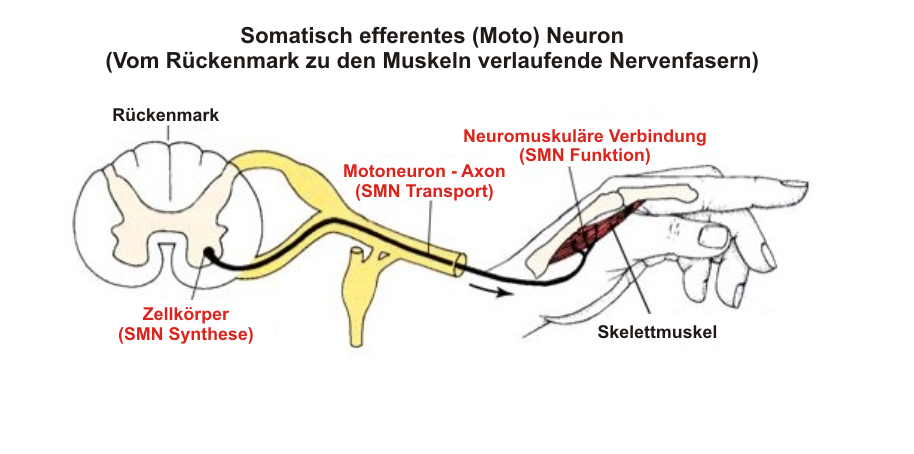
    Funktionell werden die peripheren Nerven (also die Axone von den Nervenkernen ausgehend zum Endorgan/Muskel führend), Endplatten und Muskeln zum 2. MN gerechnet, daher manchmal auch peripheres MN genannt, obwohl das so formuliert nicht ganz korrekt ist.
    Und auch wenn die Vorderhornzellen im Rückenmark, die Hirnnervenkerne im Gehirn liegen und damit anatomisch dem ZNS angehören, werden funktionell Läsionen der Vorderhornzellen und der Hirnnervenkerne selbst ebenso wie deren Axone als periphere Läsionen oder Läsionen des zweiten MN mit konsekutiver Muskelatrophie und Hypotonie bezeichnet - im Gegensatz zu den zentralen Läsionen im Motorkortex und der weißen Substanz des Rückemmarks und Gehirns (Pyramidenbahn) mit Reflexenthemmung und abnormer Steuerung der Muskeln.

    Kommentar


      #47
      Würde man hier in T1 oder T2 gewichteten MRT auch was sehen, bei entsprechender Ausprägung? Vermutlich ggf schon?
      Vermutlich als erstes in der FLAIR-Sequenz, eben im Sinne einer hyperintensen supratentoriellen CST, insb. im Bereich der inneren Kapsel, dann auch in der normalen T2-Sequenz (aber geringer ausgeprägt).

      Kommentar


        #48
        Noch einmal etwas zum ursprünglichen Thema. @LF: wenn du ALS-Bildgebung diskutieren willst, mache dafür bitte einen neuen Thread auf.
        MR-Spektroskopie ist ja durchaus noch für Mito relevant, aber der Rest nicht.

        spectrum of myopathic findings in 50 patients with the 3243A>G mutation

        "...Mutation heteroplasmy in the muscle varied from 30 to 94% (mean heteroplasmy 69 ± 14%), but no difference was found between the myopathic and non-myopathic patients or between those with mild and moderate myopathy...
        ...Muscle histology was abnormal in 26 out of 36 patients (72%). Myopathic findings were seen in nine of these (25%), atrophic changes in eight (22%), fat infiltration in three (8%) and inflammatory changes in one (3%). RRFs were absent in 34% of the muscle samples, few in 43%, moderate in 9% and numerous in 14%. The presence of numerous RRFs did not correlate with the severity of myopathy, but it did correlate with features such as diabetes mellitus (P = 0.001), epilepsy (P = 0.006) and lactic acidosis (P = 0.001). The mean proportion of COX-negative fibres in the muscle of the 16 patients that were analysed was 1.8% (range 0–5.0), and a significant difference in this proportion was found between the patients without myopathy and those with myopathy regardless of its severity...
        We found clinically defined myopathy in 25 out of 50 patients identified in a population-based screening as possessing the 3243A>G mutation. Most of the patients had mild or moderate limb weakness, while others had ptosis and/or other muscle symptoms. The patients with moderate limb weakness considered myopathy a major symptom. In the cases of limb involvement, the characteristic clinical features consisted of symmetric proximal myopathy, and a waddling gait was observed in most of these patients as a result of weakness in the hip abductors..
        ...The more COX-negative fibres were present in muscle histochemistry, the more severe was the myopathy clinically, whereas the number of RRFs did not predict the severity of myopathy,...
        ...This finding is probably due to the fact that the enzyme defect reflects the myopathic process more accurately than does mitochondrial proliferation...
        We found that CK was mildly elevated only in one-third of the patients and that their CK values did not correlate with the severity of myopathy, indicating that fibre necrosis or increased sarcolemmal permeability are not common in patients with 3243A>G. ...


        MYOPATHY AND PERIPHERALNEUROPATHY ASSOCIATEDWITH THE 3243A>G MUTATIONIN MITOCHONDRIAL DNA

        ...Peripheral neuropathy is not uncommon among patients with mitochondrial disease(Gemignani et al. 1982, Mizusawa et al. 1988, Eymard et al. 1991, Chu et al. 1997, Zanssen et al. 1998), and it varies in severity, from asymptomatic to moderate. Involvement isusually mild, with a chronic course. Electrophysiological studies have shown axonal neuropathy (Yannikas et al. 1986, Pezeshkpour et al. 1987, Mizusawa et al. 1991), anddemyelinating (Goebel et a1. 1986, Uncini et al. 1994, Rusanen et al. 1995) or mixedtypes of neuropathy (Molnar et al. 1996, Sciacco et al. 2001) have been observed in somepatients. These results have been confirmed by histopathological studies on sural nervebiopsies (Yiannakas et al. 1986). Furthermore, abnormalities detected by thermal threshold tests have suggested that small-diameter nerve fibres are preferentially affected (Sueet al. 1997).The aetiology of peripheral neuropathy in mitochondrial diseases is not fully known.The mutant load in the peripheral nerves has been observed to be similar to that in othertissues such as skeletal muscle (Chu et al. 1997, Fadic et al. 1997). Abnormal mitochondria in Schwann cells suggest that dysfunction in these cells may lead to demyelination orsecondary axonal damage (Schröder & Sommer 1991), while an increase in mitochondriain the vasa nervorum shows that mitochondria play a significant role in the pathogenesisof peripheral mitochondrial neuropathy (Molnar et al. 1995).Mitochondrial polyneuropathy is usually of a sensory rather than a motor type. Milddistal weakness may be present, but proximal or diffuse weakness is a sign of coexistentmyopathy (Peyronnard et al. 1980). Autonomic neuropathy has been reported as a rarefeature in mitochondrial disease (Enzi et al. 1985, Zelnik et al. 1996). Polyneuropathy is amajor feature in the syndrome of multiple symmetric lipomatosis (Enzi et al. 1985), andradiculopathy has been reported in a case with possible KSS (Groothuis et al. 1980) and acase with acanthocytosis (Mukoyama et al. 1986)....

        ...Exertional fatigue or intolerance is a cardinal feature of impaired oxidative phosphorylation in mitochondrial myopathies, and may be the sole manifestation of an mtDNA mutation (Andreu et al. 1999a) orin a part of multisystem mitochondrial disorder (Wibrand et al. 2001). Exercise intolerance or limb weakness can be manifested in several generations (Heiman-Patterson et al.1997) and has been reported to be present in 23% of patients with defined mitochondrialdisease (Petty et al. 1986). 50Mitochondrial myopathy has been reported in patients with mitochondrial deletions(Johnston et al. 1995), single duplications (Manfredi et al. 1995), depletions (Campos etal. 1998, Moraes et al. 1989, Moraes et al. 1991, Tritschler et al. 1992), a variety of single point mutations in mtDNA (Sweeney et al. 1993, Ionasescu et al. 1994, Weber et el.1997, Hadjigeorgiou et al. 1999, Pulkes et al. 2000, Nishigaki et al. 2002), of which somemay cause late-onset myopathy (Shibasaki et al. 1973, Johnston et al. 1995, Silvestri etal. 1998), and multiple point mutations (Andreu et al. 1999b). Sporadic mutations inpolypeptide-encoding genes, particularly in the cytochrome b gene, have been identifiedin patients with isolated exercise intolerance (Andreu et al. 1998b, Andreu et al. 1999c).The myopathy associated with mitochondrial disease is usually mild proximal andsymmetric. It may occur alone, but in most cases it is part of a syndrome. Exertionalweakness or myalgias with exertional headache or nausea have been linked to suspectedmitochondrial disease (Morgan-Hughes et al. 1990). Atypical presentations of mitochondrial myopathy can also involve subacute myalgia and elevated serum CK (Cohen et al.1998), or acute respiratory failure due to respiratory muscle weakness (Barohn et al.1990, Cros et al. 1992). An iatrogenic form of mtDNA depletion is zidovudine (antiviraldrug) myopathy with RRFs (Dalakas et al. 1990, Arnaudo et al. 1991)...

        ...Muscle specimens were obtained from the anterior tibial muscle or vastus lateralis of thequadriceps muscle under local anaesthesia. The specimens were frozen in isopentanechilled with liquid nitrogen and stored at –70°C. The frozen muscle specimens were thencut into 10-µm sections and mounted on polylysine-coated slides....
        ...Seven out of 32 patients (22%; 95% confidence interval (CI), 9–40%) fulfilled the criteria for polyneuropathy in the electrophysiological examinations (I; Table 1). Mixed axonloss and demyelinating sensorimotor neuropathy was the most common type of polyneuropathy, while one patient presented with uniform demyelinating sensorimotor polyneuropathy. Polyneuropathy was more of the sensory than the motor type (I; Table 2). Theexaminations revealed a carpal tunnel syndrome (CTS) in three patients (9.4%), two ofwhom had both classic symptoms and positive provocative tests, whereas the third wasasymptomatic. One of them had diabetes mellitus as a predisposing factor, but other aetiologies of CTS were ruled out by the laboratory tests. Electrophysiological examinationdid not reveal neuropathy in 22 patients....
        ...The seven patients with electrophysiological abnormalities presented with symptoms ofneuropathy, the median value for their neuropathy symptom scores (NSS) being 5 (I;Table 3). The polyneuropathic patients showed significantly more neurological deficits inthe detailed clinical examination (neuropathy disability score, NDS) than the otherpatients, their median score being 79, while those without neuropathy had a median scoreof 3. The patients with polyneuropathy also presented with other clinical features of amitochondrial syndrome (I; Table 2). Their functional disability was considered moreserious than that among the other patients, as reflected by a significant difference in theRankin scores between these groups (I; Table 3). The degree of 3243A>G mutation heteroplasmy in muscle was not correlated with the presence of neuropathy, but higher age63and male gender did increased the neuropathy risk. The frequency of diabetes mellitusdid not differ between the groups. Proximal myopathy was confidently diagnosed in sixout of the seven patients with polyneuropathy, and ragged red fibres were found in five ofthem. No fibre type grouping or other neurogenic changes in muscle histochemistry werefound among the patients with polyneuropathy....

        Kommentar


          #49
          ...Muscle histology was abnormal in 26 out of 36 patients (72%; 95% CI, 57–88%). Thefindings in the pathological samples consisted of myopathic changes in nine cases (25%),atrophic changes in eight (22%), fat infiltration in three (8%) and inflammatory changesin one (3%). The mean proportion of the COX-negative fibres in the muscle of the 16patients who were analysed was 1.8% (range 0–5.0%), and a significant difference in thisproportion was found between patients patients with and without myopathy regardless ofits severity (II; Table 2). RRFs were absent in 34% of the muscle samples, few in numberin 43%, moderate in 9% and numerous in 14%. Most of the RRFs were COX-positive.The presence of numerous RRFs did not correlate with the clinical severity of myopathy,but it did correlated with features such as diabetes mellitus (p = 0.001), epilepsy (p =0.006) and lactic acidosis (p = 0.001). Ultrastructural examination of the muscle samplesrevealed that variations in mitochondrial size and shape were common among patientswith limb weakness (II; Figure 2). Furthermore, both type I and type II crystals werepresent more frequently in these patients....

          Kommentar


            #50
            Im Prinzip wäre doch interessant, ob man mittels Bildgebung eine ALS Mimics infolge einer Mito von einer ALS unterscheiden kann, wenn man schon in der Röhre ist, kann man ja mehr Einstellungen / Sequenzen mitlaufen lassen und entsprechend Befunden lassen. Kann man natürlich auch im eigenen Thread diskutieren.

            Hab hier mal einen Thread dbzgl gemacht:

            Zuletzt geändert von letzte Frage; 31.01.2019, 20:23.

            Kommentar


              #51
              "ist der erwähnte Veränderte Soffwechsel im Bild 3, des PDF."

              Bei Bild 3 geht es nicht um veränderten Stoffwechsel.
              It's a terrible knowing what this world is about

              Kommentar


                #52
                Zitat von letzte Frage Beitrag anzeigen
                Also müsste dann ja beim Isaac Syndrom die NLG bei deutlicher Atrophie auffällig sein, da die Bündel der MN einen Nerv bilden...?
                Ein Nerv besteht nicht aus einem Bündel von MN. Ein einzelnes MN ist ein Nerv.
                It's a terrible knowing what this world is about

                Kommentar


                  #53
                  Zitat von letzte Frage Beitrag anzeigen
                  Voxel mit signifikant reduzierter Fraktioneller Anisotropie (grün) bei ALSPatienten verglichen mit gesunden Kontrollen. Die statistischen Ergebnisse(2 sample t-test) wurden überlagert auf ein T1-gewichtetes Bild. Die Hauptdiffusionsrichtung ist gelb dargestellt.A: Coronar-Schicht mit FA-Abnahme bilateral unterhalb des Motorund Prämotorkortex sowie im Corpus callosum.B: Coronar-Schicht mit FA-Abnahme bilateral in der Pyramidenbahnsowie im Corpus callosum.C: Axial-Schicht mit FA-Abnahme bilateral im hinteren Schenkel derCapsula interna.


                  Würde man hier in T1 oder T2 gewichteten MRT auch was sehen, bei entsprechender Ausprägung? Vermutlich ggf schon?
                  Ist die Frage, ob das nur bei ALS so ist oder nicht auch bei anderen, ähnlichen Erkrankungen.
                  It's a terrible knowing what this world is about

                  Kommentar


                    #54
                    Bei einer neurogenen Atrophie versorgt infolge der Reinnervation ein MN über dessen Axon eine Gruppe von Muskelfasern desselben Fasertyps. Ansonsten ein einzelnes MN eine Muskelfaser.
                    Zuletzt geändert von pelztier86; 31.01.2019, 21:18.

                    Kommentar


                      #55
                      die Frage, ob das nur bei ALS so ist oder nicht auch bei anderen, ähnlichen Erkrankungen.
                      das ist sicherlich der Fall. Es geht hier wohl auch eher um Grundlagenforschung, und die ist sicherlich nicht geeignet für die klinisch-diagnostische Anwendung noch dazu bei einem anderen Krankheitsbild.

                      Kommentar


                        #56
                        Ein MN-Kern (oder -Kopf) versorgt immer nur die zu ihm gehörenden Axone. Axone liegen ja nicht in der Gegend rum und warten darauf, dass ein MN-Kern kommt.
                        Bei der Reinnervation wird die Verbindung des Muskels zu einem Axon des zuständigen MN wiederhergestellt. Erwischt der Muskel dabei einen anderen Nerv kommt es zur Dyskinesie.

                        Sehe gerade, dass Du den Fehler oder Verschreiber schon selber bemerkt und den Beitrag korrigiert hast.
                        Zuletzt geändert von KlausB; 31.01.2019, 21:24.
                        It's a terrible knowing what this world is about

                        Kommentar


                          #57
                          @Klaus
                          ich wollte es erst ein wenig anders beschreiben, und dann kam zunächst das Kuddelmuddel heraus, das ich aber dann gleich bemerkt habe...

                          Kommentar


                            #58
                            Das hatte ich dann auch so ähnlich vermutet und daher meinem Beitrag den Kommentar zugefügt.

                            Habe meinen Beitrag da aber so stehen gelassen, rein zur allgemeinen Information.
                            It's a terrible knowing what this world is about

                            Kommentar


                              #59
                              Zitat von KlausB Beitrag anzeigen
                              Ein Nerv besteht nicht aus einem Bündel von MN. Ein einzelnes MN ist ein Nerv.
                              Steht in meinem Link anders:

                              Beachten Sie, dass viele Axone (schwarz) zusammengebündelt einen Nerv bilden (gelb).


                              Der Autor scheint jetzt nicht gerade unseriös:

                              Dr. Hans S. Keirstead ist Privatdozent für Anatomie und Neurobiologie am Reeve-Irvine Research Center, University of California in Irvine, U.S.A.,

                              Kommentar


                                #60
                                Zitat von pelztier86 Beitrag anzeigen
                                Kann man den Faden mal antaggen? Das geht in anderen Foren auch....
                                und es wäre schade, wenn ich mir die Mühe schon gemacht habe, wenn der Faden untergeht, wenn er nicht mehr aktuell ist...
                                Also wenn es dir wichtig ist, dann schreibs ins Wiki rein, dann wirds auch ernstgenommen von Leuten die explizit danach suchen. Ist mir gerade deshalb eingefallen, da ich mal ein Medi offlabel verordnet bekommen habe, nachdem ich den offlabel gebrauch ins Wiki reinschrieb. ^^ Wohl gemerkt wird das Medikament natürlich offlabel dazu verwendet, das wusste der Arzt nur nicht, aber als ers im Wiki las gings klar... Ärzte schauen nunmal, oft auch Wissenschaftler (kaum ein normaler - deutscher User - wird sich das durchlesen) ins Wiki und von da dann meist die Quellen.
                                Zuletzt geändert von letzte Frage; 01.02.2019, 02:17.

                                Kommentar

                                Lädt...
                                X